

Plato's CAVE - Comupterized Augmented Virtualization Environment

2D CT scan image
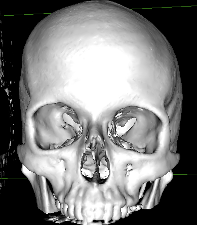
3D reconstruction
Two dimensional imaging, such as the standard cat scan (CT scan) or magnetic resonance imaging (MRI) has been around since 1972. It was invented by British engineer Godfrey Hounsfield of EMI Laboratories, England, and independently by South African born physicist Allan Cormack of Tufts University, Massachusetts. Since that time, although the resolution of the images have improved, it remains relatively the same technology. Doctors continue to use the "slices" shown on 2 dimensional CT scans to educate medical students, surgical residents, and even patients. As the images are in 2D, it can be quite disorienting even to medical residents, let alone patients who are trying to learn about their own pathology. As such, we are making push to bring 3D technology into the main stream. It is a sad state of affairs when one considers that the technology in video games are now surpassing what we are utilizing in medicine. The change from a 2D Pong game to that of 3D worlds such as Mario 3D or Call of Duty came quickly due to improved realism and depth perception. Utilizing virtual imaging centers such as Plato's CAVE, we are able to turn 2D imaging into 3D pictures, videos, and holograms. We also have the capability to use 3D printers to print out patient anatomy along with the disease so that we can carefully study the anatomy of the intricate blood vessels and nerves around certain disease processes such as benign tumors or cancers. We can import these images directly into a 3D virtual simulator so that we can surgically prepare for the actual surgery. And just as importantly, we can show these images in 3D to patients which enables them to understand their pathology in a language that they are more familiar with. In order to prove this concept works, we have published articles on this subject such as this, which shows that 3D helps immensely in improving anatomy understanding. We are also working on publishing an article shown here which looks at improving patient's understanding of their disease process and surgery to improve the patient surgical consent process. The results so far have been predictably very positive. Clearly, the technology is already here to make the 2D to 3D transition. In fact, we are also utilizing Plato's CAVE technology to now push the envelope of 4D (time-motion) and 5D (flow dynamics) imaging. Although the pace of medical development continues at a blistering pace, some aspects of utilizing the technology is not making its way into educating medical students, residents, and patients. This is due to physicians and educators being comfortable working in 2D. Like stated earlier, 2D CT scans have been around since the early 1970's and doctors have trained and used them consistently since then. Sometimes it is difficult to break tradition. I see this trend shift to 3D slowly happening around us. I expect in the next 5 years 3D will become mainstream - which would be a ground breaking shift similar to what we experienced when we shifted from plain x-rays to CT scans.